

CASE OF THE WEEK
2019-10 / APRIL 22
(CONTRIBUTOR: FADI BRIMO)
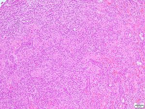
A 67-year-old male with a 2.9 cm organ-confined renal mass.


Quiz
What is the correct diagnosis?
a. Fumarate-hydrate deficient renal cell carcinoma (HLRCC-associated RCC)
b. Papillary renal cell carcinoma, type 2
c. Papillary renal cell carcinoma (biphasic squamoid variant)
d. Metanephric adenoma
e. Mucinous tubular and spindle cell carcinoma (MTSC)
1. c
Papillary renal cell carcinoma (biphasic squamoid variant)
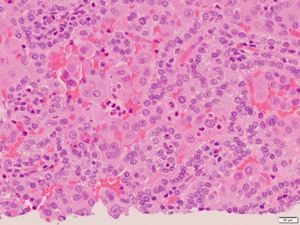
Biphasic squamoid papillary RCC is characterized by the presence of two cell populations: larger eosinophilic cells with abundant cytoplasm and higher grader nuclei forming aggregates or small nests and alveoli, and smaller cells with clear or amphophilic scant cytoplasm and lower grade nuclei. A characteristic finding is the presence of emperipolesis within the large squamoid cells, which show positivity for cyclin-D1. Otherwise, both cells populations are reactive with PAX-8, CK7, AMACR, CD10, and EMA.
Papillary renal cell carcinoma type 2 lacks the dual cell population noted herein and is composed of pseudostratified columnar cells with abundant eosinophilic cytoplasm and prominent nucleoli. Currently, papillary RCC type 2 is a diagnosis that should only be made only after having excluded other RCC subtypes such as MiT-family translocation RCC, FH-deficient RCC, and collecting duct carcinoma.
FH-deficient RCC shows a variety of architectural patterns, commonly displays papillary architecture, and is typically composed of eosinophilic cells with prominent nucleoli and peri-nucleolar halos.
Metanephric adenomas are composed of tubules and papillae lined by cuboidal cells with very scant basophilic cytoplasm in the absence of a dual cell population. In addition they are negative for CK7 and AMACR and positive for WT1 and CD57.
MTSC may show morphological and immunohistochemical overlap with biphasic squamous papillary RCC, but the dual cell population is typically lacking. In addition, MTSC typically contain mucinous and low-grade spindled areas.
Biphasic squamoid papillary RCC is not recognized as a distinct entity in the 2016 WHO classification and many of the cases previously reported in the literature as ‘solid papillary RCC with glomeruloid features’ share almost identical morphological features with it. Currently, it is still considered to be a variant that falls within the spectrum of papillary RCC, type 1 based on morphological, immunohistochemical and molecular similarities. Those tumors have been shown to be frequently multifocal, and even bilateral. Importantly, they are not all uniformly indolent and cases with recurrence, metastases or death from the disease are well documented.
Biphasic papillary renal cell carcinoma is a rare morphological variant with frequent multifocality: a study of 28 cases. Trpkov K, Athanazio D, Magi-Galluzzi C, Yilmaz H, Clouston D, Agaimy A, Williamson SR, Brimo F, Lopez JI, Ulamec M, Rioux-Leclercq N, Kassem M, Gupta N, Hartmann A, Leroy X, Bashir SA, Yilmaz A, Hes O.
Histopathology. 2018 Apr;72(5):777-785.
Biphasic Squamoid Alveolar Renal Cell Carcinoma: A Distinctive Subtype of Papillary Renal Cell Carcinoma? Hes O, Condom Mundo E, Peckova K, Lopez JI, Martinek P, Vanecek T, Falconieri G, Agaimy A, Davidson W, Petersson F, Bulimbasic S, Damjanov I, Jimeno M, Ulamec M, Podhola M, Sperga M, Pane Foix M, Shelekhova K, Kalusova K, Hora M, Rotterova P, Daum O, Pivovarcikova K, Michal M.
Am J Surg Pathol. 2016 May;40(5):664-75
Fadi Brimo
McGill University Health Center
fadi.brimo@mcgill.ca
Kidney
biphasic, squamoid, papillary, carcinoma