

CASE OF THE WEEK
2020-12 / MARCH 24
(CONTRIBUTOR: FADI BRIMO)
55 year old male with a 0.5 cm bladder polypoid lesion.


Quiz
1. What is the correct diagnosis?
a. Nephrogenic adenoma
b. Cystitis cystica et glandularis
c. Prostatic adenocarcinoma
d. Carcinoid tumor
1. d
1. Carcinoid tumor
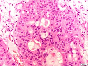
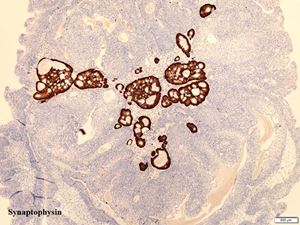
The pictures show a proliferation of urothelial cells typical of Von Brunn nests and cystitis cystica et glandularis. Admixed within are tubular and cribriform structures lined by uniform eosinophilic cells with delicate chromatin, bland cytological features and no mitoses. On immunohistochemistry, the latter population was positive for synaptophysin and chromogranin and negative for CK7 and GATA3 confirming the diagnosis of carcinoid.
Carcinoids of the bladder are rare lesions, usually small in size (mean size is 5 mm) and show a typical tubular and cribriform architectures (pseudoglandular). Other described architectural patterns are the nested, cord-like and trabecular growth patterns, which have been shown to be associated with a potentially more aggressive behavior. As illustrated in this example, the association with cystitis cystica et glandularis is frequent, which may lead to the carcinoid tumor being overlooked and the lesion being considered non-neoplastic. The tumors are typically located in the trigone or bladder neck areas and usually present with hematuria or irritative symptoms. Interestingly, carcinoid syndrome has not been reported in bladder carcinoid unlike other organs. Most carcinoid tumors are small, limited to the lamina propria and have an excellent prognosis following transurethral resection. Rare cases of large, deeply invasive tumors with more aggressive behavior have however been described. One should be aware of the fact that carcinoid-like areas can be seen in small cell carcinomas of the bladder, in which case the proper classification is small cell carcinoma.
Nephrogenic adenoma typically displays a papillary in addition to the tubular component, lacks cribriform architecture and has tubules with an atrophic appearance. Cells are positive with PAX8 and negative with neuroendocrine markers.
Prostatic adenocarcinomas involving the bladder are typically large in size, show prominent nucleoli and are positive for prostatic markers (NKX3.1, PSA, PSMA, Prostein).
Chen, YB, Epstein JI. Primary carcinoid tumors of the urinary bladder and prostatic urethra: a clinicopathologic study of 6 cases.
Am J Surg Pathol. 2011 Mar;35(3):442-6.
Fadi Brimo
McGill University Health Center, Montreal, Canada
fadi.brimo@mcgill.ca
Bladder
carcinoid