CASE OF THE MONTH
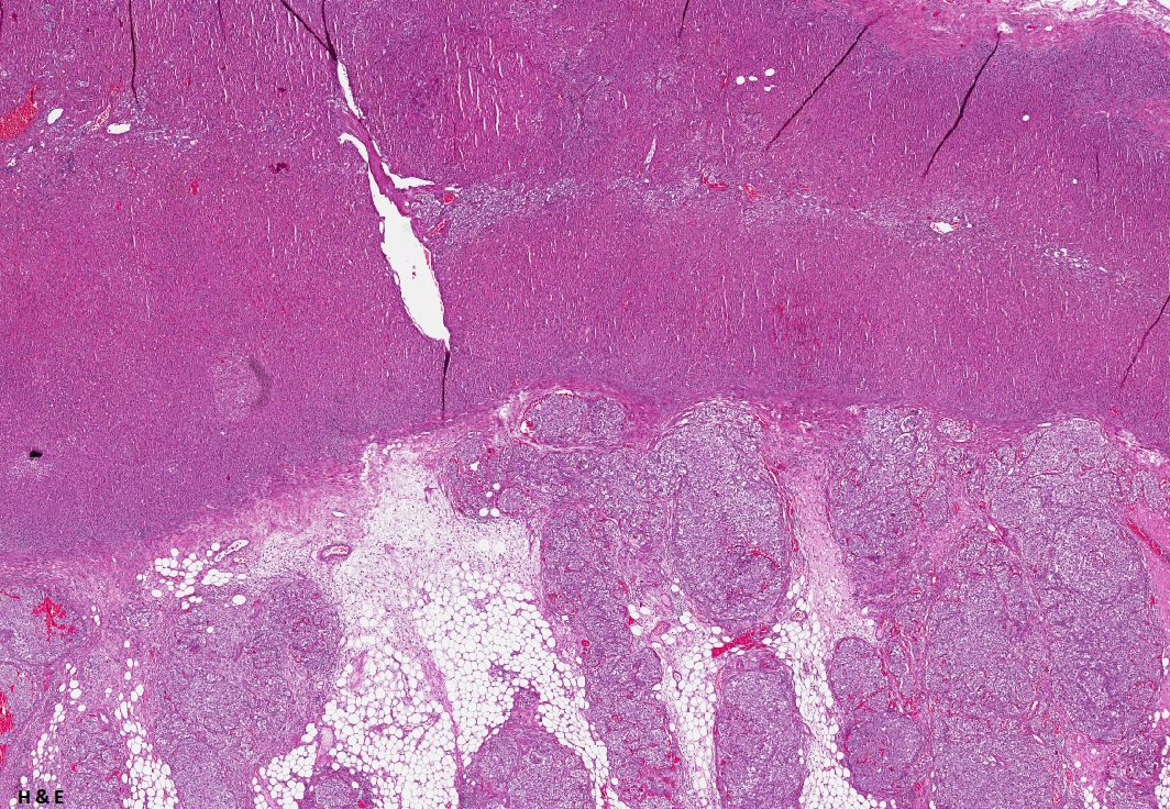
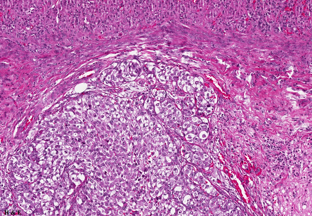
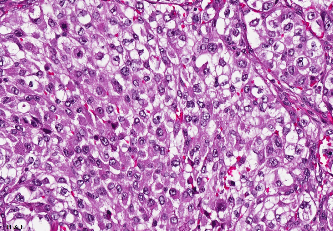
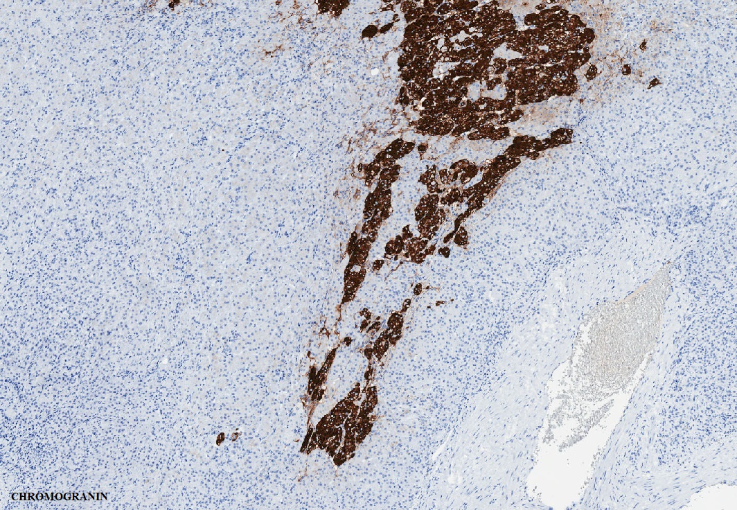
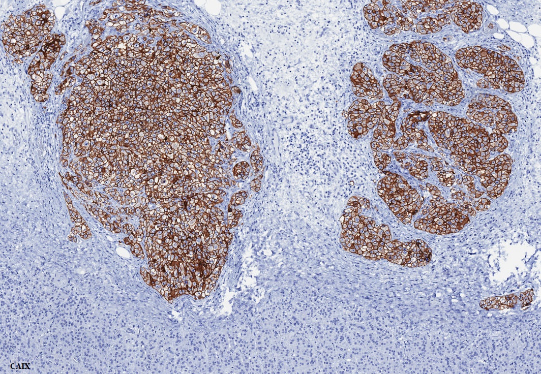
A 65-year-old man presents with a 5 cm mass in the left adrenal gland. He has a prior history of a renal cell tumor (though specific details are not available)
Contributors :
Ekta Jain, MD, PostDoctoral Clinical Research Fellow, The University of Alabama at Birmingham, Alabama, USA
Rodolfo Monitorini, MD, IFCAP, Molecular Medicine and Cell Therapy Foundation, Università Politecnica delle Marche, Ancona, Italy
Cristina Magi-Galluzzi, MD, PhD, FCAP, Professor & Chair of Pathology, The University of Alabama at Birmingham, Alabama, USA
What is the most likely diagnosis?
a. Adrenal cortical carcinoma, clear cell variant
b. Pheochromocytoma with aberrant CAIX expression
c. Secondary deposit of clear cell renal cell carcinoma, ISUP grade 3
d. Metastatic melanoma
e. Perivascular epithelioid cell tumor (PEComa)
Which immunohistochemical profile would most support a diagnosis of metastatic clear cell RCC in this adrenal mass?
a. Positive for PAX8, CAIX, and CD10; negative for SF1 and inhibin
b. Positive for SF1 and inhibin; negative for PAX8
c. Diffuse chromogranin and synaptophysin positivity
d. Positive for HMB-45 and Melan-A; negative for CAIX
e. Positive for TFE3 and cathepsin K
1. Correct answer is c.
2. Correct answer is a.
Secondary deposit of clear cell renal cell carcinoma, ISUP grade 3
On histologic examination, the adrenal lesion was composed of solid nests of tumor cells with prominent nucleoli visible at low-power magnification and cytoplasm that was partly clear and partly eosinophilic. Based on this morphology, the initial impression favored a primary adrenal neoplasm, prompting immunohistochemical workup including chromogranin. However, the chromogranin staining highlighted the normal adrenal medulla rather than the tumor, leading to a reassessment. Additional immunohistochemistry, including CAIX, was performed and proved critical in establishing the final diagnosis.
Although renal cell carcinoma (RCC) is known to metastasize to nearly any organ, adrenal involvement is relatively uncommon, despite close anatomic proximity. Adrenal metastases are often incidental findings on imaging and typically clinically silent. This case highlights the diagnostic challenge of evaluating an adrenal mass in a patient with a remote history of renal tumor. The histologic features—solid nests of cells with prominent nucleoli and partly clear, partly eosinophilic cytoplasm—are classic for clear cell RCC. Initial interpretation favored a primary adrenal tumor, but misleading chromogranin positivity in the native adrenal medulla emphasized the importance of correlating IHC findings with histology and distribution. Subsequent CAIX positivity, along with supportive morphology, confirmed the lesion as a metastatic clear cell RCC (ISUP grade 3). This case underscores the need to consider metastatic RCC in adrenal lesions, particularly in patients with a prior history of renal malignancy, and highlights the diagnostic utility of markers such as CAIX, PAX8, and CD10, when interpreted in the appropriate histologic and clinical context in resolving diagnostic uncertainty.
1. Ochsner A, Malshy K, Golijanin B, Mega A, Collins S, Maxwell A, Khaleel S, Jay B, Golijanin D. Adrenal Metastasis Ablation in Oligometastatic Renal Cell Carcinoma: A Case Series. Clin Genitourin Cancer. 2025 Apr;23(2).102295.
2. Li H, Hes O, MacLennan GT, Eastwood DC, Iczkowski KA. Immunohistochemical distinction of metastases of renal cell carcinoma to the adrenal from primary adrenal nodules, including oncocytic tumor. Virchows Arch. 2015 May;466(5):581-8.
Ekta Jain, MD, PostDoctoral Clinical Research Fellow, The University of Alabama at Birmingham, Alabama, USA
Rodolfo Monitorini, MD, IFCAP, Molecular Medicine and Cell Therapy Foundation, Università Politecnica delle Marche, Ancona, Italy
Cristina Magi-Galluzzi, MD, PhD, FCAP, Professor & Chair of Pathology, The University of Alabama at Birmingham, Alabama, USA
Adrenal gland
Metastatic renal cell carcinoma, adrenal mass, CAIX, chromogranin