CASE OF THE MONTH
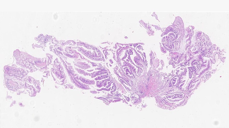
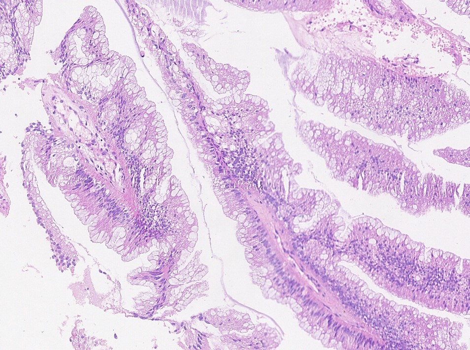
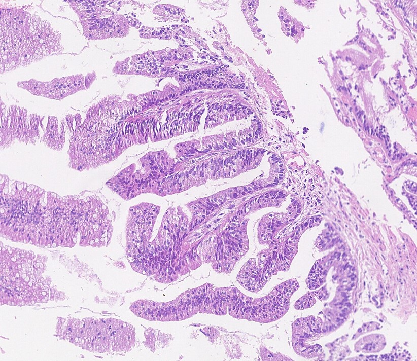
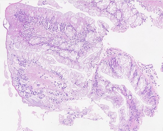
A female patient in her 80s underwent surgical removal a bladder lesion.
Contributors :
Maiara Ferreira de Souza
Oncoclinicas Precision Medicine
Recife, Pernambuco, Brazil
Luiza Barbosa Oliveira
Oncoclinicas Precision Medicine
Salvador, Bahia, Brazil
Daniel Athanazio
Oncoclinicas Precision Medicine
Federal University of Bahia
Salvador, Bahia, Brazil
What is the correct diagnosis?
Papillary urothelial carcinoma
Villous Adenoma
Mucin-rich Traditional Serrated Adenoma
Bladder Adenocarcinoma
Cystitis cystica et glandularis with intestinal metaplasia
Mucin-rich Traditional Serrated Adenoma
The urothelium shows a remarkable plasticity and – as a consequence – urothelial carcinoma may show a broad range of divergent differentiations and subtypes. In addition, the non-neoplastic urothelium may undergo different metaplastic changes including squamous differentiation, glandular differention (cystitis glandularis) and nephrogenic adenoma / metaplasia.
Cystitis glandularis may be associated with extensive intestinal metaplasia. Intestinal metaplasia is a potential precursor of intestinal-type neoplasms such villous adenoma, adenocarcinoma in situ and invasive adenocarcinoma. These neoplasms usually show intestinal phenotype. Serrated lesions – commonly seen in the colon – are rare in the bladder. There is one well documented case of Traditional serrated adenoma (TSA) and one case of hyperplastic (hypermucinous) polyp in the bladder. Updated WHO criteria categorize serrated lesions into hyperplastic polyp, sessile serrated lesion, and TSA. Hyperplastic polyp is the most common lesion and makes up 75% of serrated polyps. Sessile serrated lesion is defined by the presence of at least one aberrant crypt – and diagnostic architectural changes include horizontal growth along the muscularis mucosae, dilation of the crypt base or serrations extending into the crypt base. TSA is the least common serrated lesion in the colon and diagnosed based on the observation of slit-like clefted serration, abundant eosinophilic cytoplasm with bland pencillate nuclei, and ectopic crypt formation. Most TSAs contain only scattered goblet cells, although a mucin/goblet cell–rich TSA has been described.
AbdullGaffar B, Keloth T, Bagheri F. A Serrated Hyperplastic Colonic Polyp of the Urinary Bladder. Urology. 2019 Jan;123:e4-e6.
Pai RK, Rosty C, Makinen M, et al. Colorectal serrated lesions and polyps. In: WHO Classification of Tumours Editorial Board. Digestive system tumours [Internet]. Lyon (France): International Agency for Research on Cancer; 2019 (WHO classification of tumours series, 5th ed.; vol. 1). Available from: https://tumourclassification.iarc.who.int/chapters/31.
N Kalimuthu S, Serra S, Hafezi-Bakhtiari S, Colling R, Wang LM, Chetty R. Mucin-rich variant of traditional serrated adenoma: a distinct morphological variant. Histopathology. 2017 Aug;71(2):208-216.
Özsoy Ş, Deniz K, Temiz MZ, Semerciöz A. Traditional Serrated Adenoma of the Urinary Bladder: The First Report. Int J Surg Pathol. 2023 Aug;31(5):638-645. doi: 10.1177/10668969221113495.
Maiara Ferreira de Souza
Oncoclinicas Precision Medicine
Recife, Pernambuco, Brazil
Luiza Barbosa Oliveira
Oncoclinicas Precision Medicine
Salvador, Bahia, Brazil
Daniel Athanazio
Oncoclinicas Precision Medicine
Federal University of Bahia
Salvador, Bahia, Brazil
Urinary bladder
Bladder; Serrated lesions; Bladder Neoplasms.